Fertilisation in Humans
Fertilisation in Humans
Fertilisation is the union of male and female gametes to form a zygote. This happens in all sexually reproducing organisms. We need to study about human fertilisation in detail.
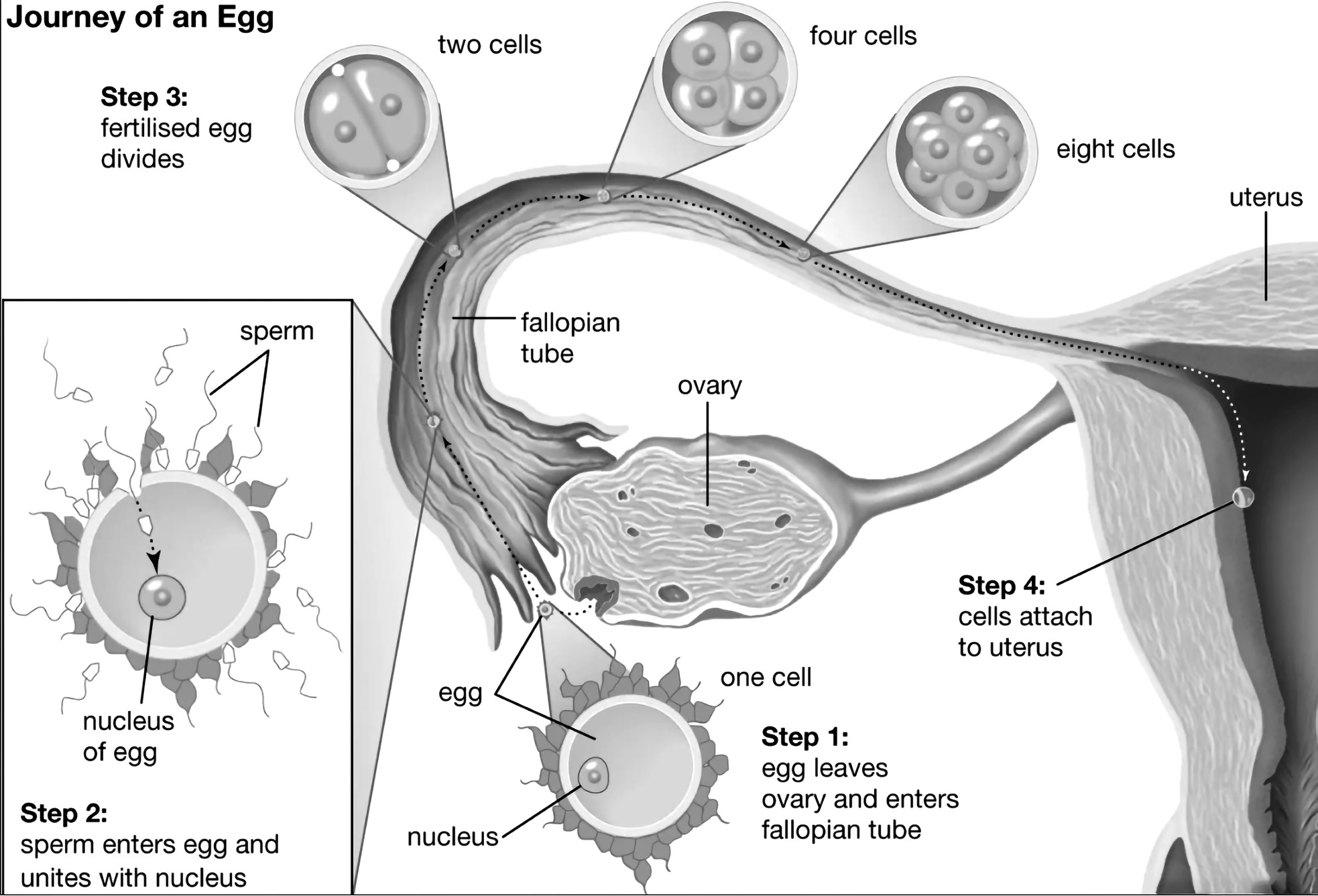
When the human sperm reaches the boundary of the female ovum, it starts a reaction called as the acrosomal reaction to allow the passage of its nucleus through the selectively permeable membrane of the female ovum.
What is important to remember here is that the entire sperm does not enter the ovum, only the nucleus does.
Then the nucleus goes on to fuse with the nucleus of the ovum leading to fertilisation and formation of the zygote

Features of the Zygote
It has nuclei containing chromosomes from the parents.
It has organelles (Mitochondria, Golgi apparatus, etc.) only from the mother as only the nucleus of the sperm enters the ovum and not the complete cell.
Thus, it gets only the DNA and nucleus from the father, other organelles are the ovum or from the mother’s cell.
It is the first cell of the new baby.
Now, try and remember back to the first chapter when we read about Cell Organelles. Was there any DNA present outside the nucleus in the cell? If yes, which cell organelle had this extra DNA? Mitochondria has DNA apart from the nucleus of the cell. We get mitochondria only from the mother and not the father because only the nucleus of the sperm cell enters the ovum.
Pronucleus- when the nucleus of the mother’s and father’s cells haven’t made the nucleus but are about to, it is called the pronucleus. Remember the term Prokaryotes? ‘Pro’ meant older cells which did not have membrane bound nucleus. Similarly, Pro + Nucleus means ‘older’ form of nucleus since the real nucleus hasn’t formed yet.
As we can see from the above diagram, we are getting the DNA and chromosomes from both the mother and father but all other organelles we are getting from the mother’s cell.
What if mother’s mitochondria is defective?
The healthy development of a child cannot happen if there are any defects in the mitochondria of the mother. For ex- Leigh’s disease in children happens when mother’s mitochondria is defective. So, the only solution we have is to replace the Mitochondria of the mother. This is done through a process called as Mitochondrial Replacement Therapy or MRT. It originated as a special form of in-vitro fertilization (IVF) in which some or all of the future baby’s mitochondrial DNA (Mt DNA) comes from a third party. This technique is used in cases when mothers carry genes for mitochondrial diseases.
Reproduction and It's Types
Reproduction: Reproduction is the biological process by which new individuals, or children, are created from their parents.
Types of Reproduction
There are two methods for animals to procreate. These are the two methods of reproduction: 1. sexual and 2. asexual.
Sexual Reproduction: Animals have different reproductive organs or parts for males and females. Animal reproductive organs similarly create gametes, which combine to form zygotes. The zygote is the one that becomes a new individual. Sexual reproduction is the process of creating offspring by fusing male and female gametes.
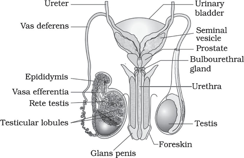
Diagrammatic view of male reproductive system (part of testis is open to show inner details
Asexual Reproduction: Asexual reproduction is the kind of reproduction in which only one parent participates.
Hydra produce new individuals from buds. We refer to this asexual reproductive process as budding.
Amoeba divides itself into two in order to reproduce. We refer to this kind of asexual reproduction as binary fission.
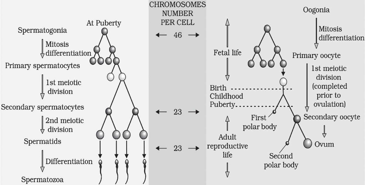
Humans are sexually reproducing and viviparous (development of the zygote happens within the body and not outside in an egg). The male reproductive system is composed of a pair of testes, the male sex accessory ducts and the accessory glands and external genitalia. Each testis has about 250 compartments called testicular lobules, and each lobule contains one to three highly coiled seminiferous tubules. Each seminiferous tubule is lined inside by spermatogonia and Sertoli cells. The spermatogonia undergo meiotic divisions leading to sperm formation, while Sertoli cells provide nutrition to the dividing germ cells. The Leydig cells outside the seminiferous tubules, synthesise and secrete testicular hormones called androgens. The male external genitalia is called penis.
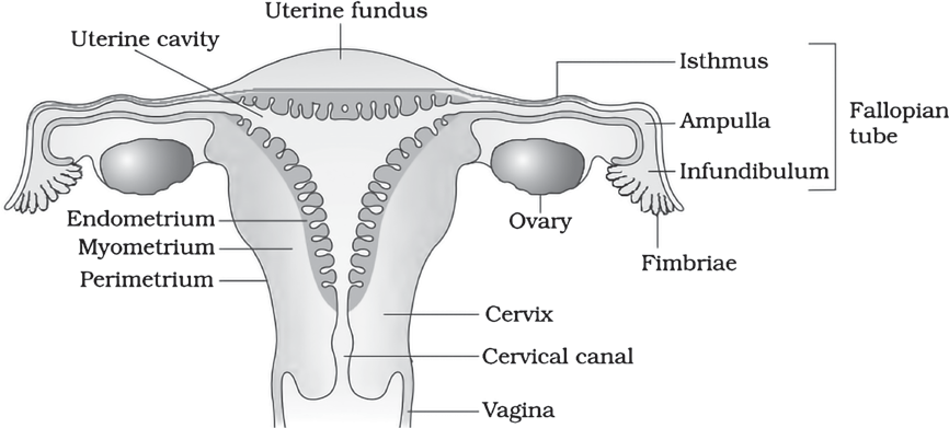
Diagrammatic sectional view of the female reproductive system
The female reproductive system consists of a pair of ovaries, a pair of oviducts, a uterus, a vagina, external genitalia, and a pair of mammary glands. The ovaries produce the female gamete (ovum) and some steroid hormones (ovarian hormones). Ovarian follicles in different stages of development are embedded in the stroma. The oviducts, uterus and vagina are female accessory ducts. The uterus has three layers namely perimetrium, myometrium and endometrium. The female external genitalia includes: mons pubis, labia majora, labia minora, hymen and clitoris. The mammary glands are one of the female secondary sexual characteristics. Spermatogenesis results in the formation of sperms that are transported by the male sex accessory ducts. A normal human sperm is composed of a head, neck, a middle piece and tail. The process of formation of mature female gametes is called oogenesis. The reproductive cycle of female primates is called menstrual cycle. Menstrual cycle starts only after attaining sexual maturation (puberty). During ovulation only one ovum is released per menstrual cycle. The cyclical changes in the ovary and the uterus during menstrual cycle are induced by changes in the levels of pituitary and ovarian hormones. After coitus, sperms are transported to the junction of the isthmus and ampulla, where the sperm fertilises, the ovum leading to formation of a diploid zygote. The presence of X or Y chromosome in the sperm determines the sex of the embryo. The zygote undergoes repeated mitotic division to form a blastocyst, which is implanted in the uterus resulting in pregnancy. After nine months of pregnancy, the fully developed foetus is ready for delivery. The process of childbirth is called parturition which is induced by a complex neuroendocrine mechanism involving cortisol, estrogens and oxytocin. Mammary glands differentiate during pregnancy and secrete milk after child-birth. The new-born baby is fed milk by the mother (lactation) during the initial few months of growth.
Chromosomes
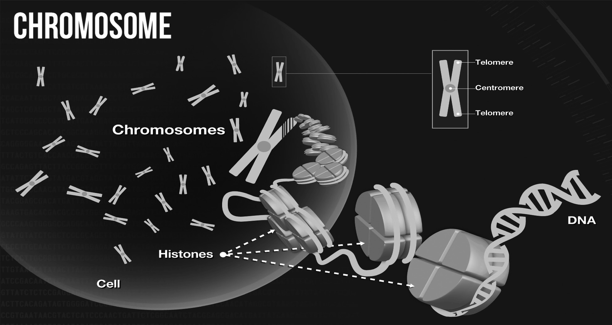
What are Chromosomes?
In the last chapter we read about DNA coiling. When DNA is coiled around histones, they form chromatin. When coiled further, supercoiled DNA forms chromosomes. The scale of this supercoiling of DNA is so huge that if a chromosome is unravelled into individual DNA molecules, the length would be nearly 2 metres! Chromosomes exist in pairs (2*n).
The term chromosome comes from Greek words for colour- chroma and body- soma. Scientists gave this name because chromosomes are stained strongly by certain dyes used in research.
What do Chromosomes do?
The unique structure of chromosomes keeps DNA tightly wrapped around spool-like proteins, called histones. As we have read in the last chapter, for an organism to grow and function properly, cells must constantly divide to produce new cells to replace old, worn-out cells. During cell division, it is essential that DNA remains intact and evenly distributed among cells. Chromosomes are a key part of the process that ensures DNA is accurately copied and distributed in the vast majority of cell divisions. Still, mistakes do occur on rare occasions.
Changes in the number or structure of chromosomes in new cells may lead to serious problems. For example, in humans, one type of leukaemia and some other cancers are caused by defective chromosomes made up of joined pieces of broken chromosomes.
Number of chromosomes
To understand chromosome number in different species, we need to understand what haploid and diploid mean. Haploid (half n) means half of the chromosome number is present in the cell, while diploid (full 2n) means full chromosome is present in the cell. Here, ‘n’ refers to the number of chromosomes characteristic for the species. For humans n is 23 meaning humans have 46 total and 23 pairs of chromosomes. The reason the sex cells have only half is because during reproduction, the other half comes from the sex cells of the other partner.
In asexually reproducing species, the chromosome number is always the same. But in sexually reproducing species, the chromosome number is diploid (2n) in case of body or somatic cells and haploid (n) in case of sex or germ cells.
What are somatic cells? What are germ cells?
Somatic cells: Somatic terms are derived from the word “Soma” which means “body”. They make up the whole organism, except reproductive or undifferentiated cells, like stem cells.
Features of Somatic Cells
An organism’s development and expansion are the result of its somatic cells.
They are necessary for renewal and restoration. Somatic cells have the same chromosome makeup as the organism and go through mitosis. Compared to gametes, which are created during meiosis (reduction division) and have a haploid set of chromosomes, somatic cells have a diploid set of chromosomes.
Germ Cells: The cells that allow for the sexual development of new creatures are known as germ cells. The embryo’s primordial streaks are the source of germ cells. Germ cells are used to make gametes.
Features of Germ Cells
These give rise to the gametes of a sexually reproducing entity.
In many animals, gametes originate primitively and pass through the intestine of the embryo to the developing gonads. Germ cells are visible in the gonads—the testes in males and the ovaries in females.
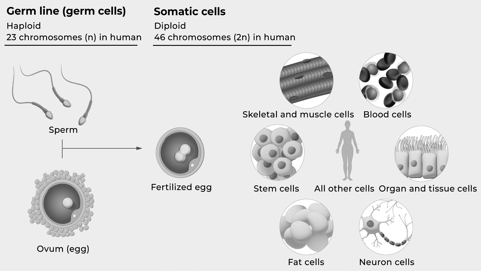
Why are there haploid cells? If chromosomes exist as pairs why do haploid cells have only half?
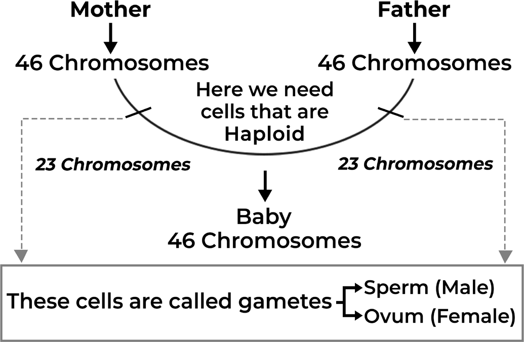
Haploid cells are needed because an offspring gets half the chromosome from each parent. Both mother and father have 23 pairs of chromosomes (total 46) which has to pass to the offspring. So, each parent passes half their chromosomes so that the offspring also has 23 pairs of chromosomes. Thus, we need a cell which only has 23 chromosomes without any pairs. This is the objective of haploid cells or germ cells or gametes. Male gametes in humans are sperm and female gametes are ovum. This is also the reason why your DNA is similar to that of your parents but never the same. Since half of it comes from the other parent and both combine to make your DNA.
Cell Division
Cell Division- Mitosis and Meiosis
There are two types of cell division. One for growth which produces diploid cells (2n) and the other for reproduction which produces haploid cells (n). Think back to the earlier discussion and imagine sperm and ovum combine to form zygote. Since, both gametes have only half the number of chromosome, the zygote has the complete chromosome needed for growth. Now when it will divide to form more cells, will it do haploid cell production or diploid cell production?
For growth, since the number of chromosomes required are the same (2n) every time, the cell division will be to ensure the production of cells with complete number of chromosomes. That is, why every cell in your body has the same DNA and genetic information. Because all of the cells in your body can be traced back to when you were a zygote and started dividing.
When cell division happens for growth, producing the diploid cells which have the same number of chromosomes, and when it leads to the passage of complete DNA of the parent to successor cells, it is called as mitosis. (Baby you Big you)
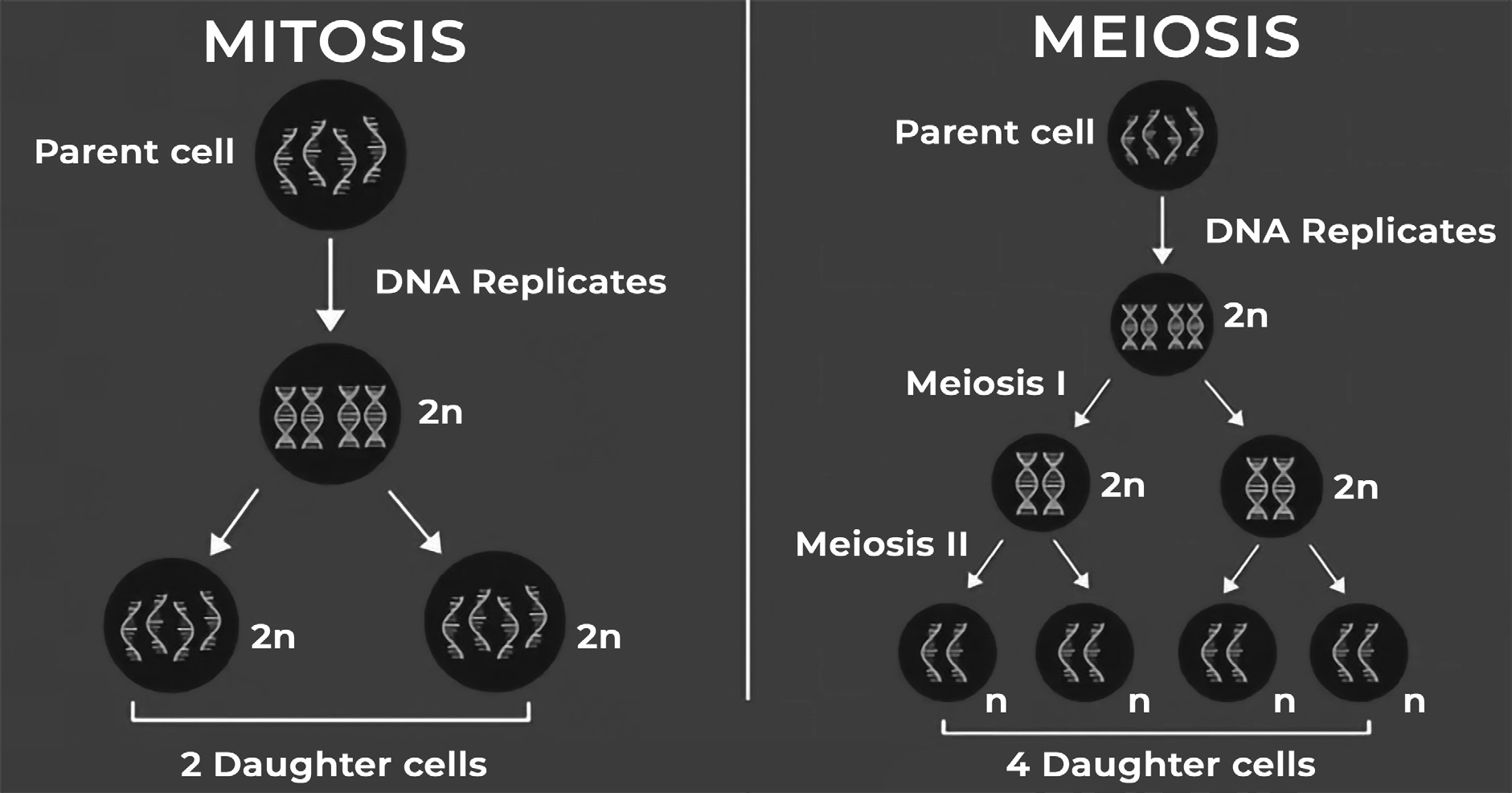
When cell division happens for reproduction, producing the haploid cells, which have half the number of chromosomes, and when it leads to the passage of only half of the DNA of the parent to successor cells, it is called as meiosis. (From your mother and father Baby you)
Ivf and Why Is It Needed
WHAT IS IVF AND WHY IS IT NEEDED?
In-vitro Fertilization (IVF) is a medical procedure in which mature egg cells are removed from a woman,fertilized with male sperm outside the body, and inserted into the uterus of the same or another woman for normal gestation woman for normal gestation.
Who needs IVF?
Male with female infertility issues.
Endometriosis
Unexplained infertility
Fertility preservation
Genetic factors
Age related infertility
Blocked fallopian tubes
Ovulatory dysfuction
Recurrent pregnancy loss(IVF/PGS-PGD)
Mrt Method-1
MRT: Method One- Pronuclear Transfer
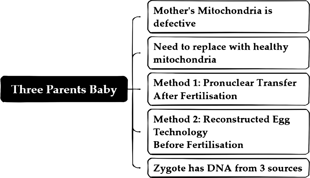
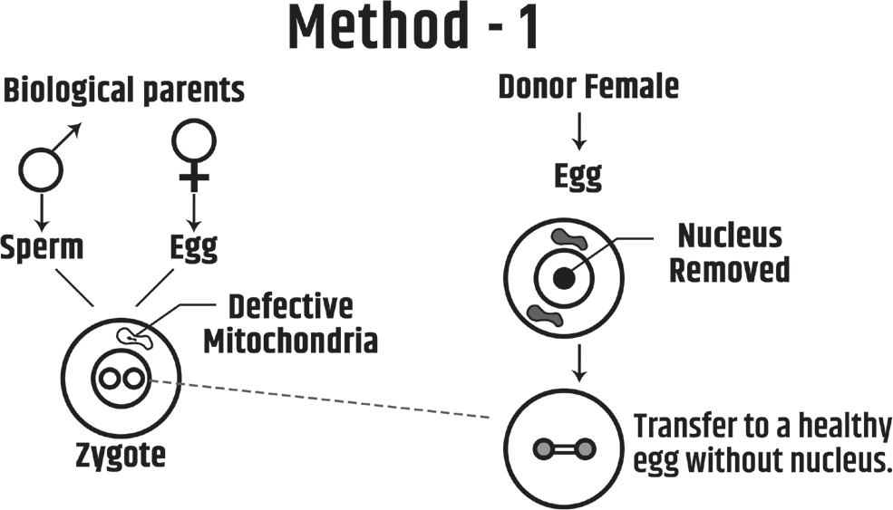
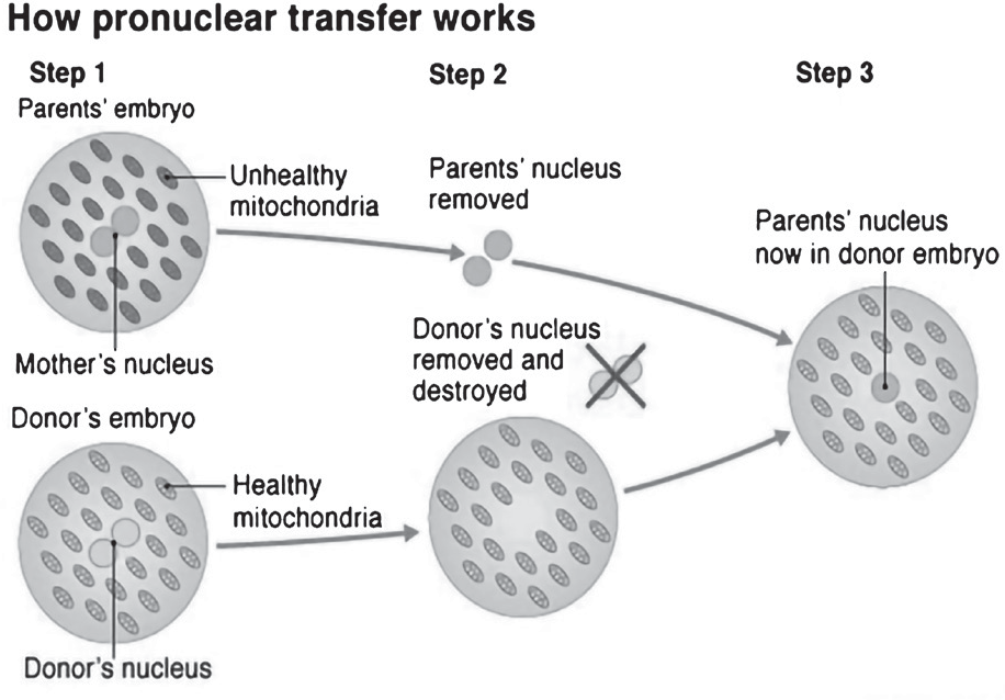
In the adjoining image, the zygote has a defective mitochondrion so MRT therapy becomes necessary in a situation like this. Ideally, we would have transferred a healthy mitochondrion from another woman and removed the defective mitochondria. But we do not have the technology for this process. So, we remove the pronucleus from the zygote and insert it into a donor female’s (who has a healthy mitochondrion with its nucleus removed) egg cell. This step is called as Pronuclear Transfer as we transferred the Pronucleus from the unhealthy zygote to a healthy egg cell of the donor. This is one way, how MRT therapy works. MRT therapy is also called three parents’ baby because the mitochondrion of the donor has some donor DNA and as a result, the baby formed would have DNA from 3 sources: nucleus and chromosomes from mother and father and mitochondrial DNA from the donor. Do note that since pronuclear formation is happening, this step happens after the fertilisation of the egg by the sperm.
Mrt Method-2
MRT: Method two- Reconstructed Egg Technology or Maternal Spindle Transfer
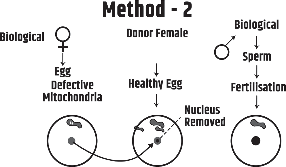
Another way to carry out MRT is before fertilisation, the nucleus from the mother’s defective egg is removed and placed in a healthy donor’s egg and then fertilised with the father’s sperm to form a healthy zygote. This process is called as Reconstructed Egg Technology or Maternal Spindle Transfer. In this case also MRT is called as three parents’ baby